 Tap to zoom
Ectopic Kidney: Causes, Symptoms, Diagnosis, and Treatment
An ectopic kidney is a kidney that is not in its usual anatomical position. Learn about symptoms, diagnosis, treatment options, and ways to manage this congenital kidney anomaly.
- Published on
- June 26, 2026
- Reading time
- 5 min read
- Last updated
- Updated: June 29, 2026
An ectopic kidney is a rare congenital condition in which one kidney is located outside its normal position. The kidney may lie near its usual site or in the pelvis, abdomen, or even the chest.
The main cause is usually a disturbance in kidney development and migration during fetal life.
Urologic surgeon Kidney stone and urinary tract specialist
Note: To improve care quality and patient satisfaction, appointments are triaged by reason for visit. Each physician sees patients within the relevant urology subspecialty.
Book appointment
An ectopic kidney may function normally, but in some cases it can lead to problems such as urinary tract obstruction, recurrent infections, kidney stones, and functional impairment.
Ectopic kidney is usually diagnosed with medical imaging such as ultrasound, CT scan, and MRI. Treatment depends on the kidney location and the patient's symptoms, and may include surgery, infection management, or other supportive measures. Correct identification and management can improve quality of life.
Ectopic kidney is thought to occur in about 1 in every 900 births, but only about 1 in 10 of these cases is diagnosed. These kidneys may be discovered while treating other conditions.
Appointments related to Urologic surgeon Kidney stone and urinary tract specialist
Public recorded-slot data is available for this destination.
Book appointment
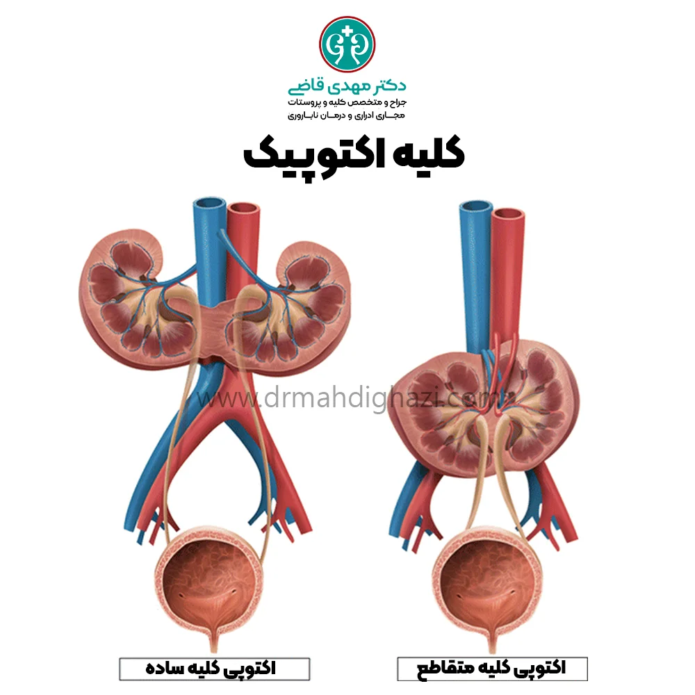
During fetal development, the kidneys bud in the lower abdomen and then ascend to their correct position. Ectopic kidneys do not move to the correct site and may be found anywhere in the abdomen, whether high, low, or on the opposite side.
Types of kidney ectopia
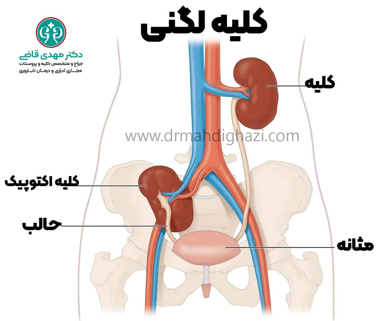
Simple kidney ectopia
This refers to a kidney that is on the correct side but in an abnormal higher or lower position, such as a pelvic kidney.
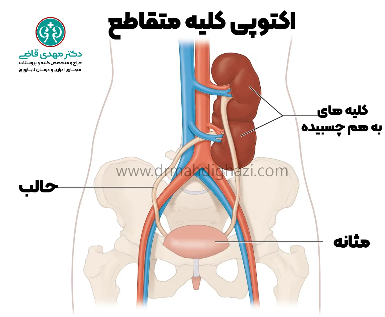
Crossed kidney ectopia
This is a condition in which one kidney crosses from its side to the other side. As a result, both kidneys are on one side of the body. They may be separate or fused. Crossed renal ectopia may also be associated with congenital anomalies of other organs.
Q&A — Urologic surgeon Kidney stone and urinary tract specialist
General questions are shown on the destination page after review.
Book appointment
How does the urinary system normally work?
The urinary system includes two kidneys, two ureters, one bladder, and one urethra. Healthy kidneys filter blood throughout the day and night. These bean-shaped organs are in the back, just below the ribs, one on each side of the spine. The kidneys are the body's main filters.
They filter about 150 liters of blood each day and produce about 1 to 2 liters of urine by removing excess water and waste from the blood.
Urine normally travels from the kidneys to the bladder and then leaves through the urethra. As filters, the kidneys also regulate many factors needed for health:
Fluid balance
Electrolyte levels, such as sodium, potassium, calcium, magnesium, and acid
Removal of waste as urine
Regulation of blood pressure and red blood cell count
Causes of ectopic kidney
This anomaly is a congenital defect with an unknown cause, but it may be related to the following:
A defect in the fetal kidney bud
A defect in the tissue that guides the kidneys to the correct location
Genetic defects
Infections, diseases, and chemicals that affect the fetus.
Symptoms of kidney ectopia
The most common symptoms associated with ectopic kidney are:
Abdominal pain
A mass in the abdomen
Blood in the urine
Complications of ectopic kidney
A kidney in an abnormal location may still work properly, but because of its changed position it may have difficulty draining urine, leading to hydronephrosis or kidney dilation.
About 1 in every 2 ectopic kidneys has some degree of obstruction.
Mild obstruction may not cause complications, but severe obstruction can have serious consequences. Over time, these obstructions may lead to other problems, such as:
Urinary tract infections (UTIs)
Kidney failure
Vesicoureteral reflux (VUR) is more common in ectopic kidneys. Reflux is a disorder in which urine flows backward from the bladder through the ureters to the kidneys.
Over time, urinary reflux can cause infection, and infection can cause permanent kidney damage.
Although a healthy, non-ectopic kidney can also develop problems such as obstruction or reflux, the likelihood of these complications is much lower in a normally positioned kidney.
Diagnosis of kidney ectopia
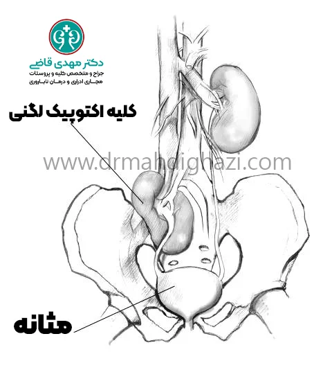
Only about 1 in 10 ectopic kidneys is diagnosed. Doctors often find ectopic kidneys while treating other conditions. They may also be found during evaluation of the symptoms mentioned earlier. The following imaging tests can help the doctor find an ectopic kidney:
Ultrasound
Intravenous pyelogram, or IVP
Voiding cystourethrogram, or VCUG image
Magnetic resonance imaging, or MRI
Pelvic kidneys can be diagnosed earlier than other ectopic kidneys.
The doctor may also request blood tests to assess kidney function.
Treatment of ectopic kidney
An ectopic kidney needs treatment only if there is significant obstruction or kidney reflux (VUR) is marked, which generally is not present in most people. If the kidney is not severely damaged or nonfunctional when the problem is discovered, surgery can relieve the obstruction or treat reflux to prevent future worsening. But if the kidney is severely scarred and does not work well, the urologic surgeon may recommend removing it.
Because vascular and ureteral abnormalities can accompany ectopic kidneys, surgery on these kidneys is challenging and requires the surgeon to have adequate familiarity and skill with the anatomy of that area.
Follow-up after treatment
If the patient has only one kidney and that kidney works well, the person can live a normal life span even after removal of the ectopic kidney.
Regular follow-up after treatment, including periodic blood tests and kidney imaging, is necessary to make sure the remaining kidney is functioning properly.
Prevention of ectopic kidney complications
An ectopic kidney may cause complications because of its abnormal position. By following certain recommendations and receiving appropriate care, serious complications can be prevented and patients' quality of life can improve.
Appropriate diet
Reduce salt intake
High salt intake can raise blood pressure and cause kidney problems. Salt intake should be limited and low-salt foods should be used.
Avoid excessive protein and oxalate intake
High protein intake, especially animal protein, can increase the kidney workload. Oxalate in some foods can also contribute to kidney stone formation. Therefore, foods containing oxalate, such as spinach, beetroot, and nuts, should be consumed in moderation.
Adequate hydration
Drink enough water
Adequate water intake helps prevent kidney stones and urinary infections. People are advised to drink at least 8 glasses of water per day. If kidney complications are present or the doctor gives different advice, the amount of water intake may change.
Hygiene care
Maintain personal hygiene
Personal hygiene and care of the genital and urinary areas can help prevent urinary infections. Regular handwashing and clean cotton underwear can be useful.
Prevent urinary infections
To help prevent urinary infections, it is better to avoid irritants such as scented soaps and hygiene products that contain chemicals.
Exercise and physical activity
Regular physical activity
Regular exercise and physical activity can help improve blood circulation and kidney function. Gentle activities such as walking, swimming, and yoga can be useful.
Regular medical follow-up
Regular visits to the doctor: regular visits are necessary to monitor the ectopic kidney and the healthy kidney. The doctor may order periodic blood tests and kidney imaging so that any change or problem is identified and treated on time.
Summary
An ectopic or displaced kidney is a rare congenital condition in which the kidney is in an abnormal location. It may be asymptomatic, but it can cause problems such as recurrent infections, urinary tract obstruction, kidney stones, and functional disorders.
Diagnosis is mainly made with imaging such as ultrasound, CT scan, and MRI. Treatment depends on kidney location and symptom severity, and may include surgery, infection management, or other supportive measures.
With correct diagnosis and management, quality of life can improve and serious complications can be prevented. An appropriate diet, adequate hydration, hygiene care, and regular medical follow-up are effective preventive measures for kidney health.
Frequently asked questions
Actions & related links
Related articles
All articlesWhat Is Polycystic Kidney Disease? (Fetal and Adult PKD)
What is polycystic kidney disease? Learn about inherited ADPKD and ARPKD, fetal and adult symptoms, complications, diagnosis, medicines, surgery, diet, fluids, and prevention-focused care.
What Is a Renal Cortical Cyst? Symptoms, Diagnosis, and Treatment
Renal cortical cyst | Simple vs. complex kidney cysts | Warning symptoms | Diagnosis and treatment | Needle drainage, laparoscopy, medication, and ablation
Kidney Transplant: Cost, Blood Type Compatibility, and Surgical Method
What is kidney transplant? Learn about cost considerations, operation duration and method, diet, who may not be eligible, success rates, isolation precautions, and post-transplant care.
What Is Pyelonephritis? Kidney Infection Symptoms, Diagnosis, and Treatment
What is pyelonephritis? Learn kidney infection symptoms in women, children, men, and pregnancy; diagnosis, antibiotics, treatment, emergency warning signs, and cystitis differences.
Comments
0 comments
No comments yet. Be the first to share your thoughts.