 Tap to zoom
Urethral Diverticulum: Symptoms and Treatment
Learn what urethral diverticulum is, its symptoms, causes, diagnosis with MRI or cystoscopy, and surgical or follow-up treatment options.
- Published on
- June 26, 2026
- Reading time
- 5 min read
- Last updated
- Updated: June 27, 2026
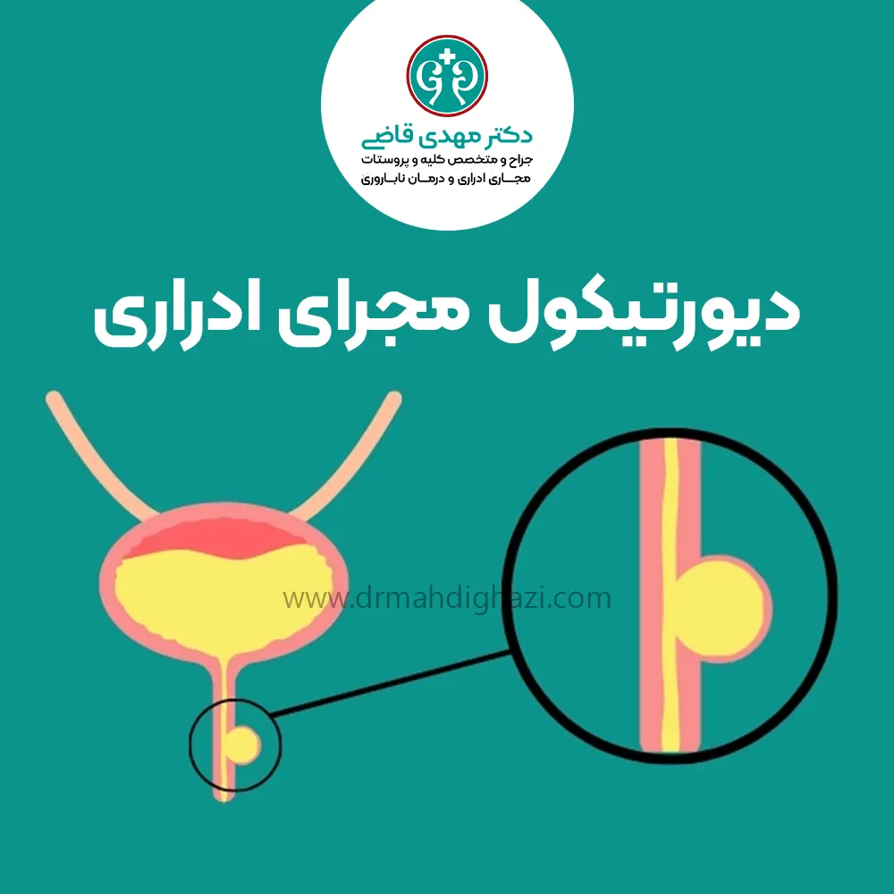
A urethral diverticulum is an abnormal sac-like structure or cavity that forms along the urethra (the tube that carries urine out of the body) and is usually connected to the urethral lumen through an opening. During urination, this sac can fill with urine, and when urine remains inside it, it can create an environment prone to recurrent infections.
A urethral diverticulum may cause different symptoms, including:
A painful lump or bulge in the vaginal wall
Chronic pain in the pelvic area
Recurrent urinary tract infections
This condition is rare and is seen more often in women between 40 and 70 years old. In general, this abnormality does not occur in children unless they have a history of surgery on the urethra.
With advances in medical imaging, diagnosis and detection of urethral diverticula have increased, and more cases are being treated. However, many cases remain untreated because they are not diagnosed or are misdiagnosed, which can lead to more serious problems and complications.
Causes of Urethral Diverticulum
The cause of urethral diverticulum is not fully understood. However, it is often associated with the following:
Recurrent infections that weaken the urethral wall.
Obstruction in glands within the urethra
Some older studies also mention congenital defects or injury during childbirth.
Symptoms
Up to 20% of patients with urethral diverticulum may have no obvious symptoms. Symptoms vary from person to person, but the most common include:
Recurrent bladder or urinary tract infections
Pelvic pain
Irritative lower urinary tract symptoms (similar to overactive bladder)
Nocturia (feeling the need to urinate several times at night)
Pain during sexual intercourse
Urinary dribbling
Blood in the urine
Vaginal discharge
Urinary retention or symptoms of urethral obstruction
Urinary incontinence
Some women feel pain or a lump in the front part of the vaginal wall. With gentle pressure, urine or pus may drain from the urethral diverticulum.
Important point: The size of a urethral diverticulum is not what matters. In some cases, a very large diverticulum may cause only mild symptoms. By contrast, a small diverticulum can cause severe pain and discomfort. Symptoms may also appear and then disappear without treatment.
Diagnostic Methods
Because urethral diverticulum does not have specific symptoms, it may be discovered during a doctor's examination or imaging.
Because detecting these diverticula can be somewhat difficult and requires relatively complex imaging, it may take years for some people to receive the correct diagnosis.
Patients with urethral diverticulum are often misdiagnosed and initially treated for other problems.
Accurate diagnosis is possible with the following:
A detailed medical history
Physical examination
Urinalysis
Cystoscopy (direct examination of the bladder and urethra using a thin lighted scope)
Imaging such as MRI or ultrasound
Examination
If a urethral diverticulum is found, the urologist may gently "milk" the sac to drain pus or urine. In women, the front part of the vaginal wall may be examined to check for a lump and pain.
Imaging
Several imaging methods can be used to find a urethral diverticulum. No test is definitely the best method. Each method has advantages and disadvantages.
The final choice often depends on the following:
Availability of the test
Cost of the test
Radiologist's expertise
Magnetic Resonance Imaging (MRI)
This type of imaging uses a magnetic field to examine the urinary tract in detail. MRI is the best method for diagnosing urethral diverticulum.
Ultrasound
This test may show a urethral diverticulum, but a follow-up MRI is needed for confirmation.
Urodynamic Studies
These tests assess the function of the lower urinary tract and may detect stress urinary incontinence (involuntary loss of urine when pressure on the bladder increases) caused by urethral diverticulum.
Video Urodynamic Studies
Because these include imaging as well, they may be able to identify the cause of stress urinary incontinence.
Treatment
In asymptomatic cases, counseling and follow-up may be sufficient, and removal of the diverticulum is not necessary if it appears low-risk. Surgery is the main treatment for urethral diverticulum. However, not every case requires surgery.
Some patients may not want surgery or may not be candidates for it.
There is not enough information about people who did not have their urethral diverticulum treated. It is unclear whether the sacs become larger or whether symptoms worsen.
Some people prefer not to take action until symptoms become more severe. In rare cases, cancer has been reported to develop in people with urethral diverticulum. If the patient does not want surgery, counseling and follow-up with a physician are very important.
Surgery for Urethral Diverticulum (Urethra)
Surgical removal of the diverticulum (diverticulectomy) is the best treatment method. This surgery should be performed by an experienced urologist who is familiar with the procedure. The diverticular sac may be connected to the urethra.
If the sac is not removed carefully, it can lead to recurrence of the diverticulum or damage the urethra. This can create the need for more complex reconstructive surgery.
Surgical Methods for Urethral Diverticulum
Surgical methods for treating urethral diverticulum include:
Incising and widening the neck of the diverticulum (performed at the point where the sac connects to the urethra)
Creating a permanent pathway from the diverticular sac into the vagina
Complete removal of the diverticulum
Other Important Considerations in Urethral Diverticulum Surgery
Complete closure and suturing of the diverticular neck (the connection to the urethra) to prevent recurrence
Complete removal of the inner lining of the diverticulum to prevent recurrence
Multilayer suturing to prevent a new tract from forming between the urethra and the vagina
If you have stress urinary incontinence (SUI), surgery to correct incontinence may be performed at the same time as diverticulum surgery.
After Treatment
If the patient decides not to have surgery, they should still see their urologist for follow-up.
If the patient decides to have surgery, however, the following points should be kept in mind.
Antibiotics are needed for at least 24 hours.
A catheter (a tube that drains urine from the bladder) is needed for 2 to 3 weeks.
Bladder spasms and occasional severe pain may occur and are managed with medication.
Two to three weeks after surgery, a voiding cystourethrogram (VCUG), an X-ray study performed during urination with contrast material, is done to check whether there is any urine leakage.
If there is no fluid leakage, the catheter is removed.
If fluid leakage is seen, voiding cystourethrography (VCUG) is repeated weekly until the leakage stops.
In most cases, leakage resolves within a few weeks.
Common Surgical Complications
Urinary tract infections
Urinary incontinence
Recurrence of urethral diverticulum
No improvement in urinary symptoms
Urethrovaginal fistula (an abnormal tract between the urethra and the vagina, which is a serious problem that requires treatment)
Recurrence of Urethral Diverticulum
If a urethral diverticulum returns, it may be for the following reasons:
The sac was not completely removed.
The connection site was not completely closed.
Dead space was left behind.
Other technical problems
Repeat surgery can be difficult. This surgery requires a high level of technical skill.
Summary
Urethral diverticulum is a rare structural abnormality of the urinary tract that forms as a sac or cavity along the urethra and connects to the urethral canal through an opening. This sac can fill with urine during urination and, as a result, create a suitable environment for bacterial growth and recurrent infections.
This condition is seen more often in middle-aged women and women over 40 and can cause symptoms such as recurrent urinary tract infections, pelvic pain, and a painful lump in the vaginal area. Although it is asymptomatic in some cases, accurate diagnosis and appropriate treatment are very important when severe symptoms occur.
Imaging methods such as MRI and ultrasound play a key role in accurate diagnosis of this disease. In severe cases, surgical removal of the diverticulum (diverticulectomy) is used as the main treatment method, while counseling and follow-up alone may be sufficient in asymptomatic cases.
However, lack of treatment or misdiagnosis can lead to complications such as recurrence and fistula formation.
After surgery, careful care and necessary follow-up, including antibiotic use, catheter use, and postoperative imaging, are essential for complete recovery and prevention of later complications.
Frequently Asked Questions
Actions & related links
Related articles
All articlesWhat Is Polycystic Kidney Disease? (Fetal and Adult PKD)
What is polycystic kidney disease? Learn about inherited ADPKD and ARPKD, fetal and adult symptoms, complications, diagnosis, medicines, surgery, diet, fluids, and prevention-focused care.
What Is a Renal Cortical Cyst? Symptoms, Diagnosis, and Treatment
Renal cortical cyst | Simple vs. complex kidney cysts | Warning symptoms | Diagnosis and treatment | Needle drainage, laparoscopy, medication, and ablation
Kidney Transplant: Cost, Blood Type Compatibility, and Surgical Method
What is kidney transplant? Learn about cost considerations, operation duration and method, diet, who may not be eligible, success rates, isolation precautions, and post-transplant care.
What Is Pyelonephritis? Kidney Infection Symptoms, Diagnosis, and Treatment
What is pyelonephritis? Learn kidney infection symptoms in women, children, men, and pregnancy; diagnosis, antibiotics, treatment, emergency warning signs, and cystitis differences.
Comments
0 comments
No comments yet. Be the first to share your thoughts.